The intricacies of the Equine placenta Anatomy: A comprehensive guide
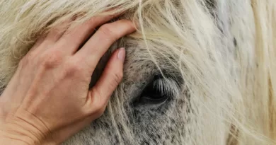
The equine placenta is an amazing organ that provides nutrients and oxygen to the fetus and removes waste products. This comprehensive guide will give you a detailed look at the anatomy of the equine placenta.
You’ll learn about the different layers, membranes, and vessels that make up this complex structure. Plus, you’ll better understand how it functions and what can go wrong. So, check out this guide whether you’re a horse owner or just interested in learning more about this fascinating structure.
What is the Equine Placenta Anatomy?
The placenta is a complex organ that develops during pregnancy to support the fetus. It is attached to the lining of the uterus. It extends into the maternal blood vessels, providing a pathway for exchanging oxygen, nutrients, and waste products between mother and fetus. The placenta also produces hormones that are essential for maintaining pregnancy.
The placenta is composed of two layers: the chorion, which is derived from the fertilized egg, and the decidua, which is derived from the lining of the uterus. The chorion contains fetal blood vessels, while the decidua contains maternal blood vessels. These two layers are separated by a thin layer of tissue called the intervillous space, through which exchanges between mother and fetus occur.
The placenta Anatomy is unfamiliar to most people, but it is something that develops during pregnancy to support the growing fetus. It comprises two layers: the chorion (from the fertilized egg) and decidua (from the uterus lining). These layers are separated by a thin layer of tissue called the intervillous space.
What does it do for the foal and mare during gestation?
The Equine Placenta is an amazing organ. It grows during the pregnancy and attaches the developing foal to the mare’s uterus. It provides nutrients and oxygen to the foal and removes waste products from its blood. The placenta also produces hormones that help to regulate the pregnancy.
During gestation, the placenta grows rapidly and becomes very thick. This helps to protect the foal from harm and provides a large surface area for exchanging nutrients and waste products between the foal and mare. The placenta also helps regulate the uterus’s temperature, keeping it at a constant temperature that is ideal for the developing foal.
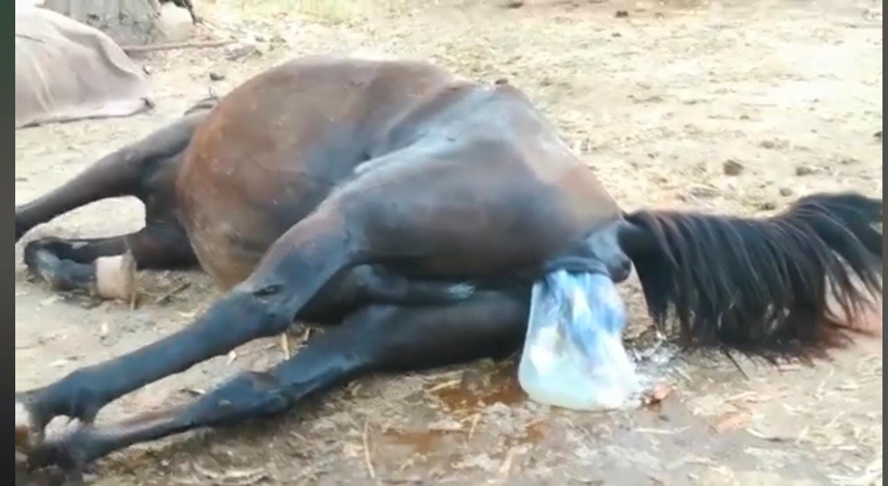
At birth, the placenta is expelled from the mare’s body and the foal’s umbilical cord. The placenta continues to provide nutrients and oxygen to the foal for a few minutes after birth until it can start breathing independently. After the placenta is expelled, it is eaten by the mare – this helps her body recover from childbirth and provides her with important nutrients to produce milk for her new foal.
How is the placenta structured, and what are its different layers responsible for fetal development and maternal health?
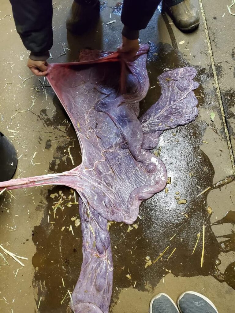
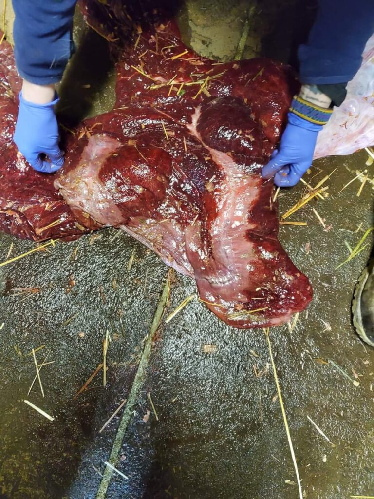
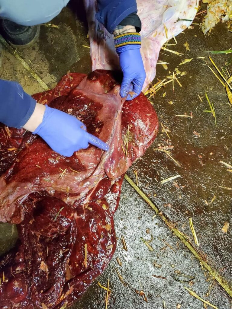
Equine placentas are classified as “cotyledonary,” meaning they have small, flattened areas called cotyledons responsible for exchanging nutrients.
There are four main layers of the equine placenta.
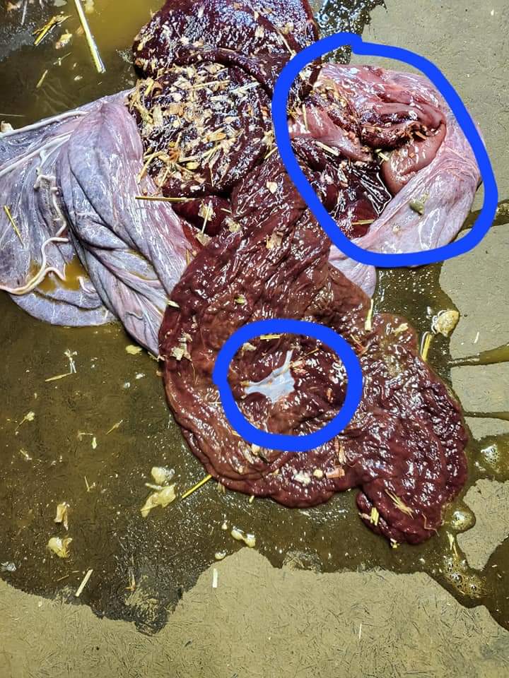
The Basal layer
The basal layer is in contact with the uterine wall and helps anchor the placenta.
The Caruncular layer
The caruncular layer is composed of connective tissue and blood vessels and helps to provide nutrients to the fetus.
The Chorionic layer
The chorionic layer is the outermost layer and helps to protect the developing fetus from infection.
The Allantoic layer
The allantoic layer contains a sac-like structure called the allantois, which stores waste products from the fetus.
The placenta is essential for fetal development and maternal health, providing nutrients, oxygen, and waste removal for the growing fetus. Without a healthy placenta, fetal development will be hindered, and maternal health may be at risk.
When does embryo implantation into the uterine wall occur, and how does this process progress over time until birth occurs?
Equine pregnancy is unique in many ways, one of which is how the embryo becomes implanted in the uterine wall. It is thought that implantation occurs on days 14-16 after fertilization, though this can vary slightly based on the individual mare.
Once implantation has occurred, the developing embryo will begin to secrete a substance known as trophoblast, which helps to attach the embryo to the uterine wall. The placenta will also start to develop at this time, and by day 28, it will be fully formed.
This process of attachment and development will continue until birth when the foal is expelled from the uterus, and the placenta will be shed. Equine pregnancy is a fascinating example of how the body adapts to support new life, and understanding the implantation process can help us better care for pregnant mares.

What common problems can affect placental function or implantation, causing complications during pregnancy or delivery?
It also serves as a physical buffer between the fetus and the mare. Common problems that can affect the placental function or implantation include:
- Reduced blood flow to the area due to changes in the mare’s vascular system caused by pregnancy
- Inflammation of the uterus is caused by infection or other conditions.
- Physical trauma to the uterus caused by foaling or other factors.
- Hormonal imbalances can interfere with normal placental function.
If not addressed, these problems can lead to complications during pregnancy or delivery, such as abortion, stillbirth, dystocia (difficulty delivering the foal), or neonatal death. Equine reproductive specialists are trained to identify and manage these conditions to help ensure a successful outcome for both mare and foal.
How has modern veterinary science improved our understanding of equine placenta and their role in successful pregnancies?
The equine placenta is the extra-embryonic membrane that connects the fetus to the uterine wall in the Equidae species. These membranes provide a structural and nutritive link between the dam and fetus and offer a barrier to potential pathogens.
Studies on the Equine placenta have been performed since the early 1800s, focusing mostly on their morphology and function. However, it was not until the mid-1900s that veterinary science began to take a closer look at equine placenta and their role in successful pregnancies. Thanks to modern technology, we now understand equine placenta anatomy and physiology better.
We can also identify placental abnormalities early on, which has helped improve pregnancy outcomes in mares. Additionally, research on the Equine placenta has led to advances in our understanding of other mammalian placentation.

Placental transfusion – what is it, why might it be necessary, and how safe is it for both mare and foal?
Equine placenta transfusion transfers Equine Placental blood from the mare to the foal. This is done by taking Equine Placental blood from the mare and transfusing it into the foal. The Equine Placental blood contains oxygen and nutrients essential for the foal’s survival.
The transfusion of Equine Placental blood into the foal helps ensure that the foal receives the oxygen and nutrients it needs to survive. Equine placenta transfusions are safe for both the mare and the foal. Equine placenta transfusions are typically only necessary when the mare cannot produce enough Equine Placental blood for the foal. When Equine placenta transfusions are necessary, they generally are only required once or twice during the foaling process.
More Articles: